
Merck Veterinary Manual, what a great reference! Musculoskeletal system, Veterinary, Merck
A baby cow is called a calf. A female calf is sometimes called a heifer calf and a male a bull calf. A heifer is a female that has not had any offspring. The term usually refers to immature females; after giving birth to her first calf, however, a heifer becomes a cow. An adult male is known as a bull.

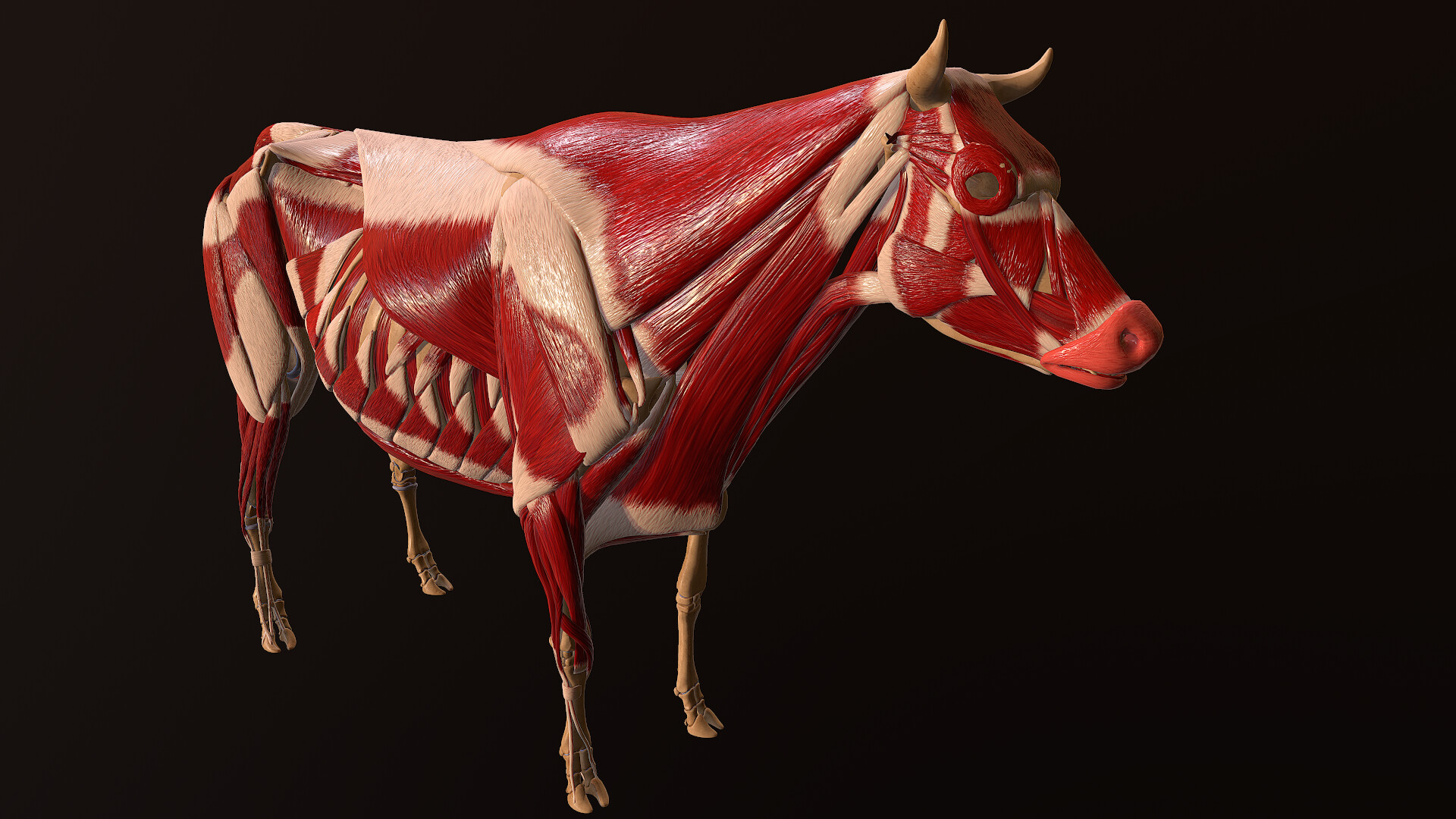
MODEL OF A COW'S ANATOMY, THE MUSCLES, FRAGONARD MUSEUM, NATIONAL VETERINARY SCHOOL OF ALFORT
The Anatomy of a Cows Stomach. Inside a cows stomach region, there are 4 digestive departments:. 1. The Rumen - this is the largest part and holds upto 50 gallons of partially digested food. This is where the 'cud' comes from. Good bacteria in the Rumen helps soften and digest the cows food and provides protein for the cow.
Muscular System Of A Cow paradetips
5 Muscles of the Forelimb 5.1 Extrinsic Musculature 5.2 Intrinsic Musculature 6 Muscles of the Shoulder 6.1 1. Lateral 6.2 2. Medial 6.3 3. Caudal (Flexors) 7 Muscles of the Elbow 7.1 Extensors 7.2 Flexors 8 Muscles of the Carpal and Digital Joints 8.1 Extensors 8.2 Flexors 9 Vasculature of the Forelimb 10 Webinars
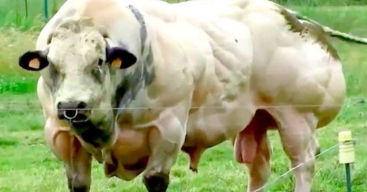
The Reason This Cow Is So Insanely Muscular The Dodo
Muscle Descriptions. Contact. Michaella Fevold, Assistant Professor of Practice Animal Science Department A213c Animal Science Building Lincoln, NE 68583-0908 (402)472-9896. [email protected]. Related Links. Beef Research; Beef Nutrition; Beef Innovations Group; Beef for Foodservice; Beef for Retail;
Cow muscles Buy Royalty Free 3D model by carlos faustino (carlosfaustino) [c33d0a1
Cow yoga pose stretches and warms up the following muscles: Hip flexors. Cow pose stretches your hip flexors, making them longer and less prone to injury. There are five muscles involved in.
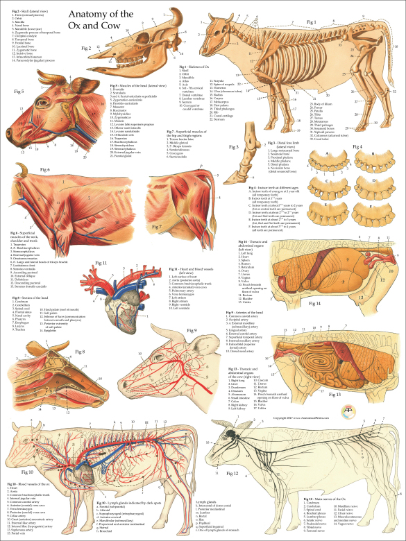
Cow Ox Anatomy Poster
The subiliac lymph nodes (bov) are found at the cranial edge of the thigh muscles, about midway between the tuber coxae and the fold of the flank. (Fig. 31.10) The paralumbar fossa (ID in bov, but also in eq) is the most common surgical site for entry into the ruminant abdomen.
ArtStation Cow anatomy sceleton muscles ligaments
Muscles of the cow's antebrachium and manus Lateral flexor muscle of cow shoulder Medial flexor muscle of cow shoulder Flexor muscles of cow elbow (arm) Extensor muscle of cow elbow (arm) Extensor muscles of cow antebrachium Flexor muscles of cow front leg anatomy Cow back leg muscles anatomy Lateral muscles of the cow hip and thigh

Muscle Groups of Cattle Diagram Quizlet
1, masseter muscle; 2, coronoid process; 3, temporal fossa; arrowheads, temporal line; 4, paracondylar process; 5, occipital condyle; 6-9 cheek teeth (Triadan numbers).. Figure 25-18 Left half of upper and right half of lower jaw of cow. Note the different shapes of the upper and lower cheek teeth and the large diastema (1).
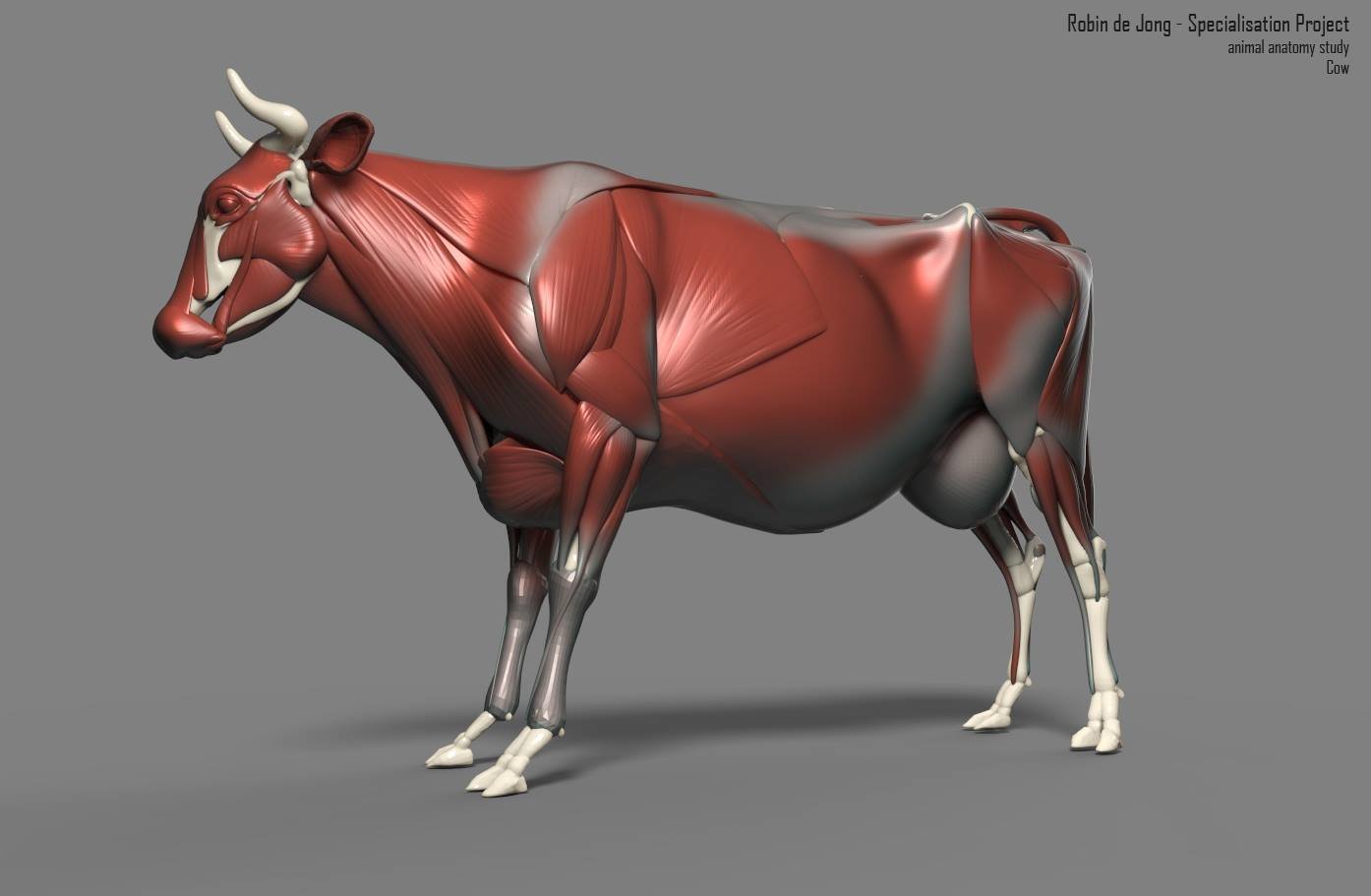
Robin de Jong cow anatomy study
Muscles of the hindlimb of a cow Cow anatomy organs Digestive organs of a cow Cow anatomy stomach Compartments of cow stomach Liver and pancreas of cow anatomy Organs of the respiratory system from a cow Lung anatomy of a cow Heart of a cow Cow hoof anatomy Cow anatomy labeled diagram Frequently asked questions on cow Conclusion Cow anatomy

Allgemeine Anatomie des Bullen und der Kuh Bildatlas
Dairy cows are judged on, and selected for, wide spread pin bones. (In the HORSE, the tuber ischii are covered by hamstring muscles.) The head of the femur, which articulates with the acetabulum, is found medially, while on the lateral side there is the greater trochanter with cranial and caudal cusps. (Figs. 4-3 and below)
.jpg)
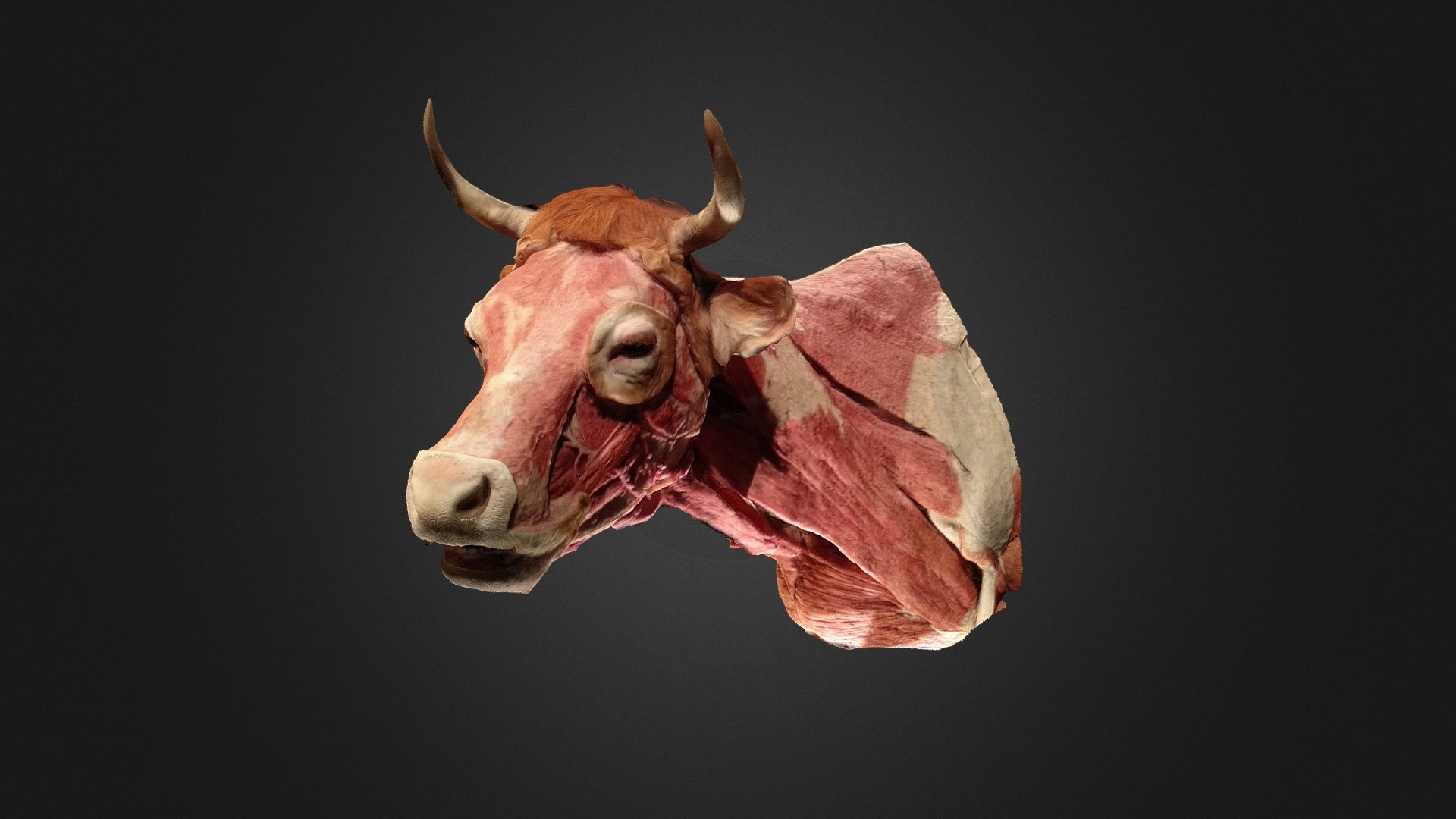
Deep muscle of cow head and neck plastinated specimen,medical teaching model, medical specimens
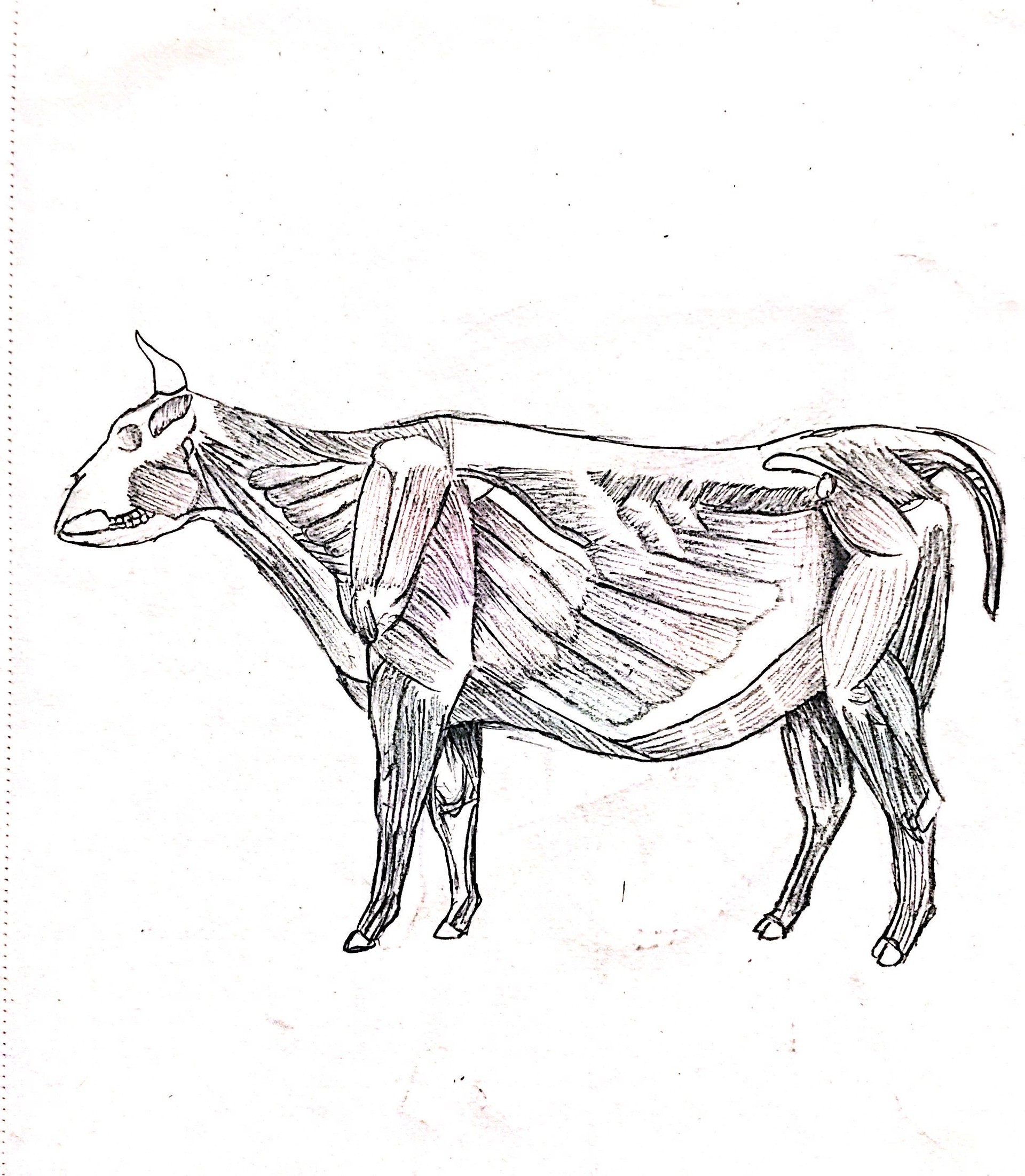
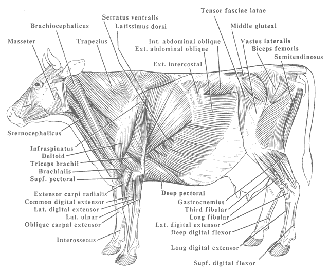
The superficial muscles of a cow are diagramed. Labels: 1, Occipito-Frontalis. 2, Orbicularis Palpaebrarum. 3, Masseter. 5, Sterno-cleido-Mastoid. 6, Trapezius. 7, Latissimus Dorsi. 8, Pectoralis. 9, 10, External and Internal oblique muscles. 11, Opening of the mammary artery and vein (milk vein). 12, Gluteii. 13, Rectus Femoris muscle.

Myology Muscles of the Pelvic Limb (COW) Diagram Quizlet
Bull-Cow - Muscles Bull-muscles Bull-Cow - Digestive system Bull-digestive systeme Bull-Cow - Sagittal section-Manus Bull-sagittal section of manus Bull-Cow - Terms of position and direction Bull-terms of position and direction ANATOMICAL PARTS Abaxial tendon Abdomen Abomasum Accessory carpal bone Acromion Adductor pollicis muscle
Anatomy
norecopa.no NORINA Bovine Anatomy: The Cow Anatomical Chart Bovine Anatomy: The Cow Anatomical Chart This chart shows views of the cow's left lateral view with the dorsal and vertebral regions indicated. Type of record: Chart/Diagram. Category: Anatomy

Bovine Cow Muscle Anatomy Poster Muscle anatomy, Large animal vet, Anatomy
Delayed treatment or unresponsiveness to treatment in cows with clinical periparturient hypocalcemia ( milk fever ), as well as calving paralysis from nerve injury after dystocia, may result in prolonged involuntary recumbency. Less common primary causes of recumbency in alert downer cows include severe hypokalemia and possibly hypophosphatemia .

Bovine Muscle Anatomy Cow Muscular System Cow muscles by uberkudzu Animals Muscular system
1 Pelvic Girdle and Hip 1.1 Bones 1.1.1 Bovine Bone Specifics 2 Joints and Synovial Structures 2.1 Sacroiliac Joint 2.2 Coxafemoral/Hip Joint 3 Musculature 4 Proximal Hindlimb including Stifle and Tarsus 4.1 Bones 4.1.1 Bovine Bone Specifics 4.2 Joints and Synovial Structures 4.3 Musculature 5 Vasculature of the Hindlimb 6 Webinars
The Superficial Muscles of a Cow ClipArt ETC
(Figure 3) The cow is very thin with no fat on ribs or in brisket and the backbone is easily visible. Some muscle depletion appears evident through the hindquarters. Figure 3. BCS 3. BCS 4. (Figure 4) The cow appears thin, with ribs easily visible and the backbone showing. The spinous processes (along the edge of the loin) are still very sharp.